Electron Microscopy Images
We have a library of images recorded over the years using our scanning and transmission electron microscopes. If you want something, ask. Few are shown below and others elsewhere. If you have questions about the images or want some specific images contact Max Guinel.
Hibiscus
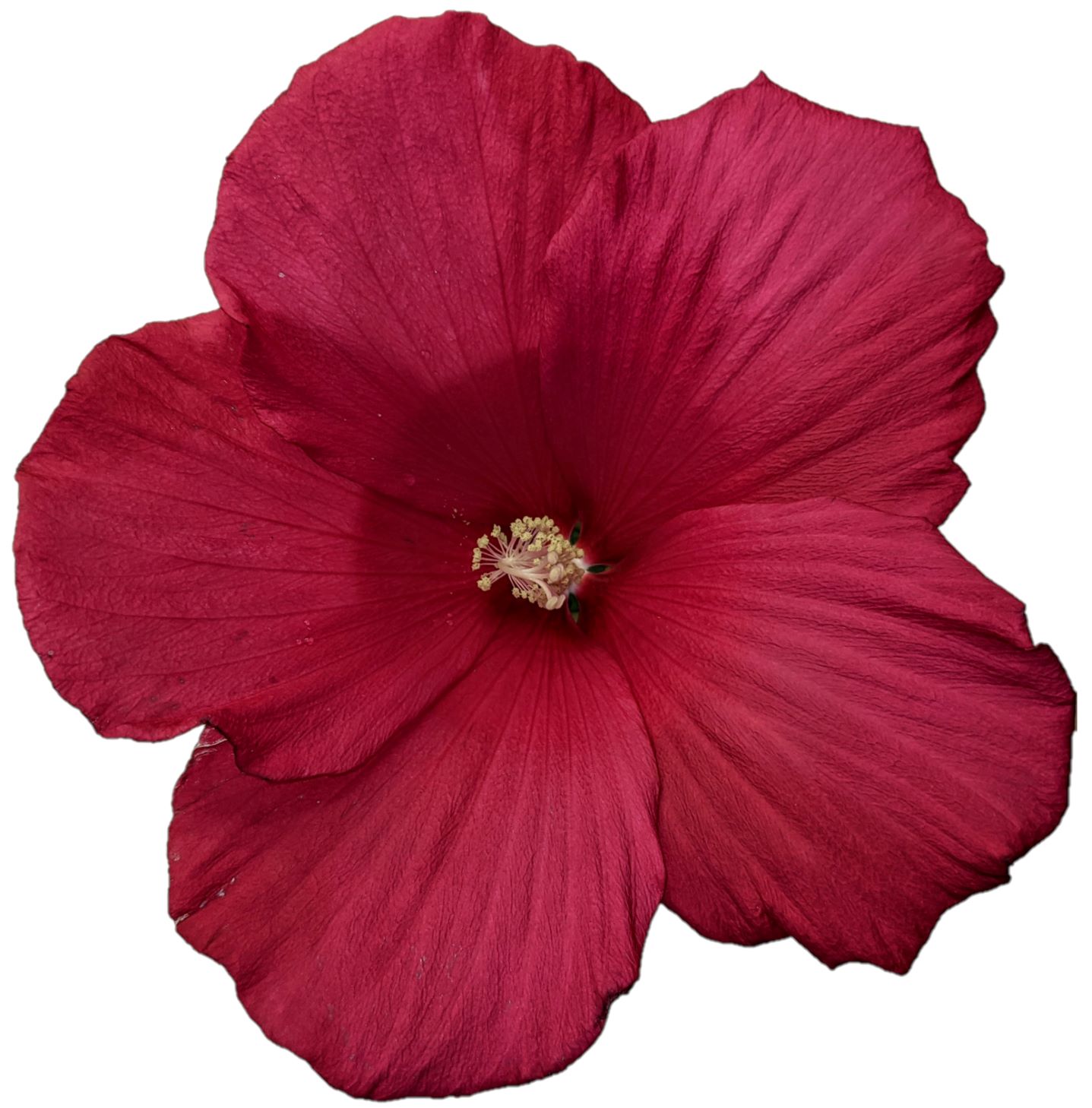
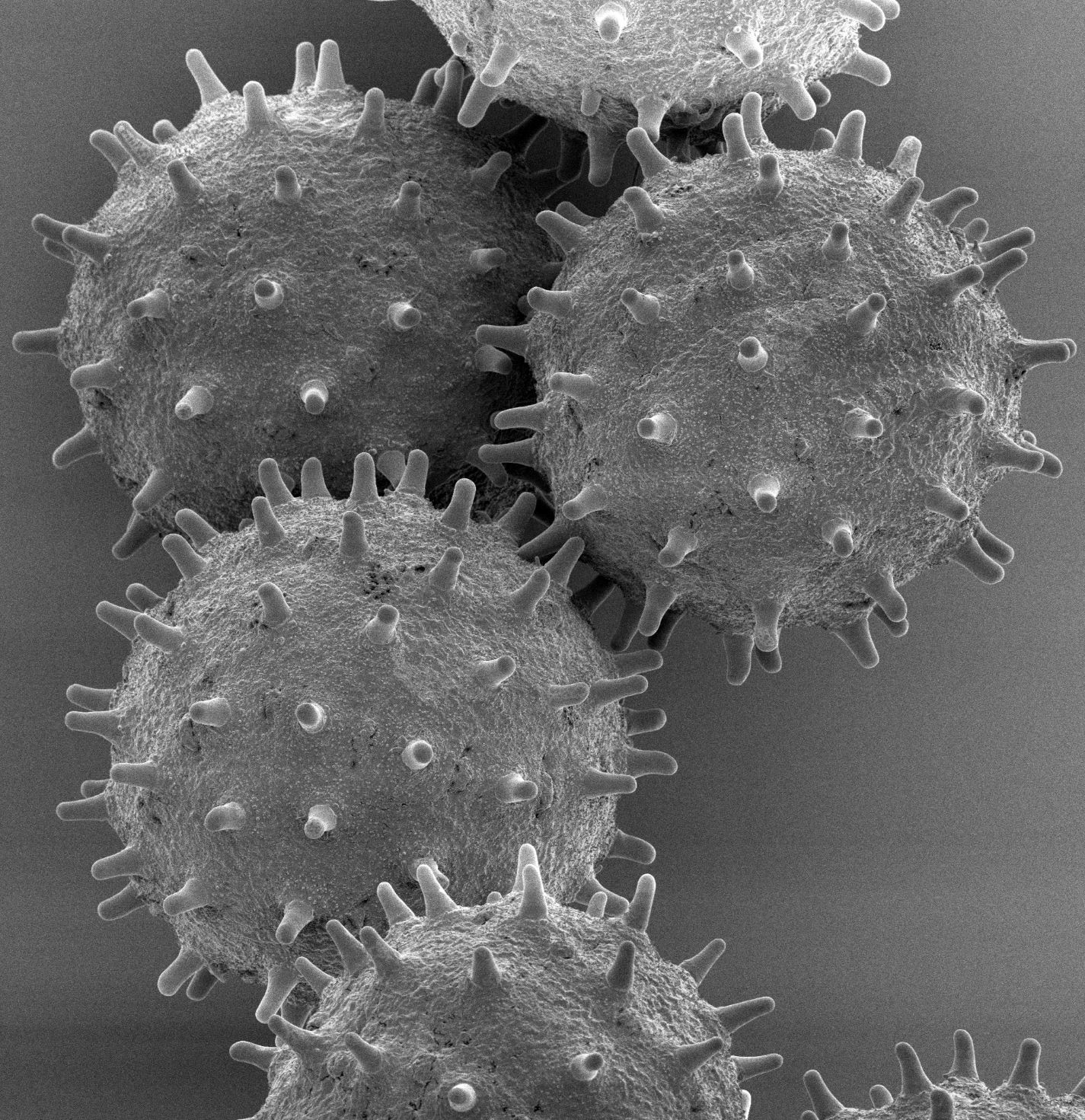
Morphy @ Dartmouth Life Sciences Greenhouse
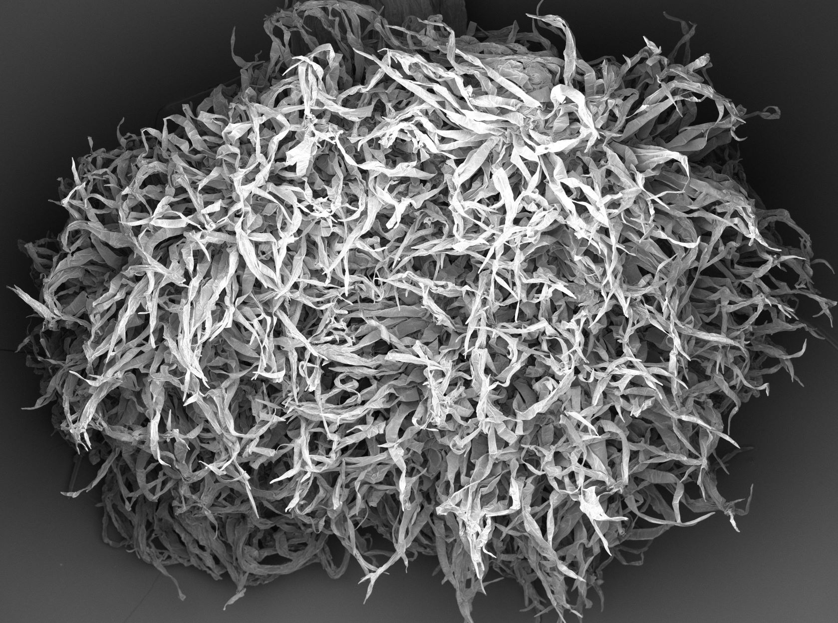
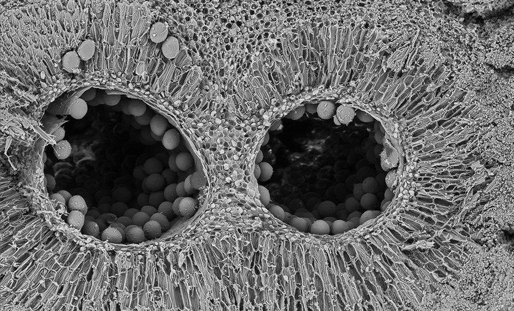
Amorphophallus titanum anther cross section. High magniification image showing pollen inside the locule (cavity where the pollen is located).
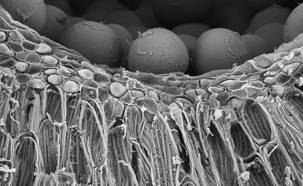 Amorphophallus titanum anther cross section, showing the locule (cavity where the
pollen is located).
Amorphophallus titanum anther cross section, showing the locule (cavity where the
pollen is located).
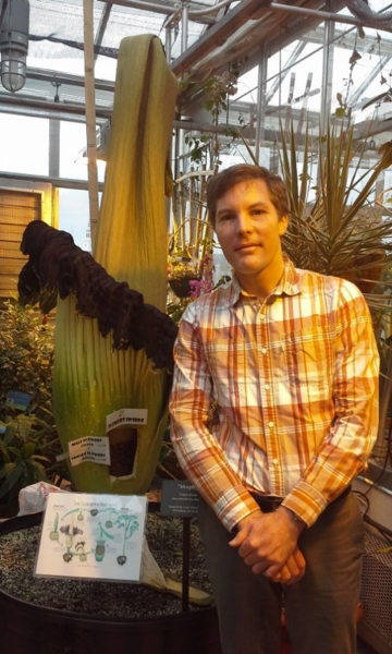 Very large flower (Max standing next to Morphy in bloom years ago).
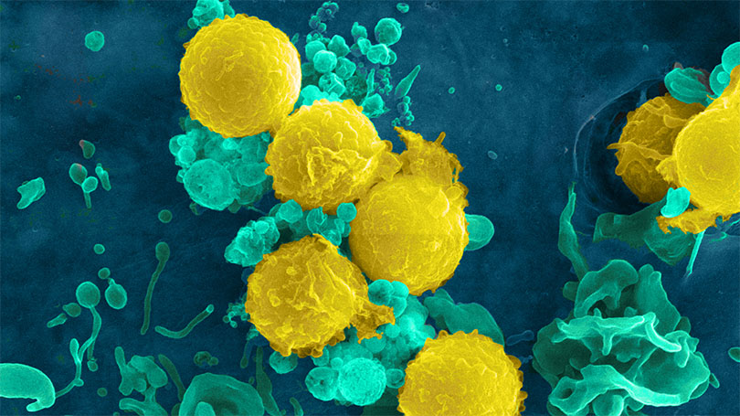
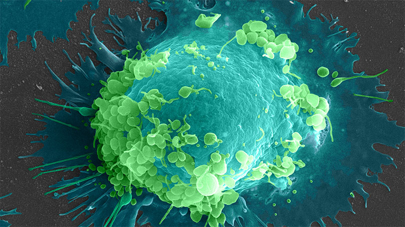
HIV Virus
Tissue culture cell line, infected with human immunodeficiency virus (HIV)
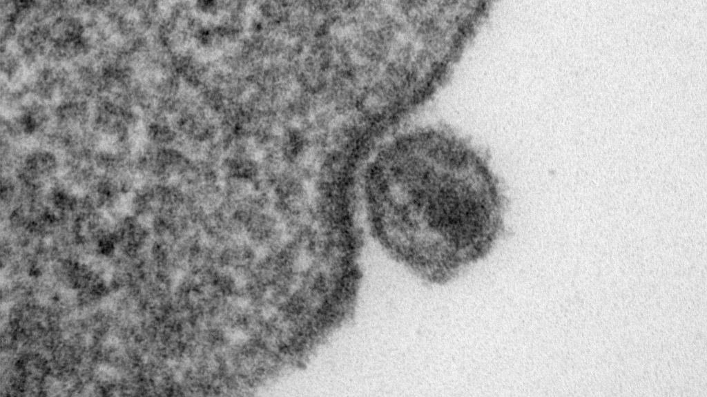 HIV particles are 90-120nm in diameter.
The process of infection: (1) virus attaches to the cell via the CD4 molecule on the surface of the cell. The CD4 molecule on the lymphocyte acts as a cell surface receptor for HIV. (2) fusion of the virus envelope with the host cell membrane. (3) nucleocapsid, containing the genetic material, is carried into cytoplasm by endocytosis. Instrument: JEOL 1010 TEM.
View a video about the HIV infection pathway.
Red Blood Cells
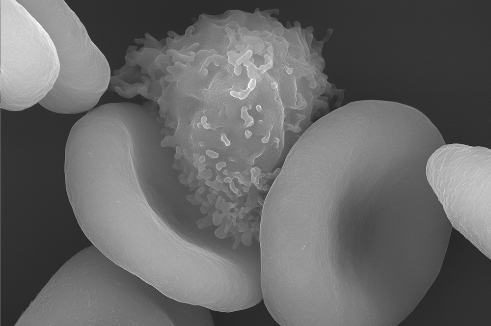 Human red blood cells and a lymphocyte.
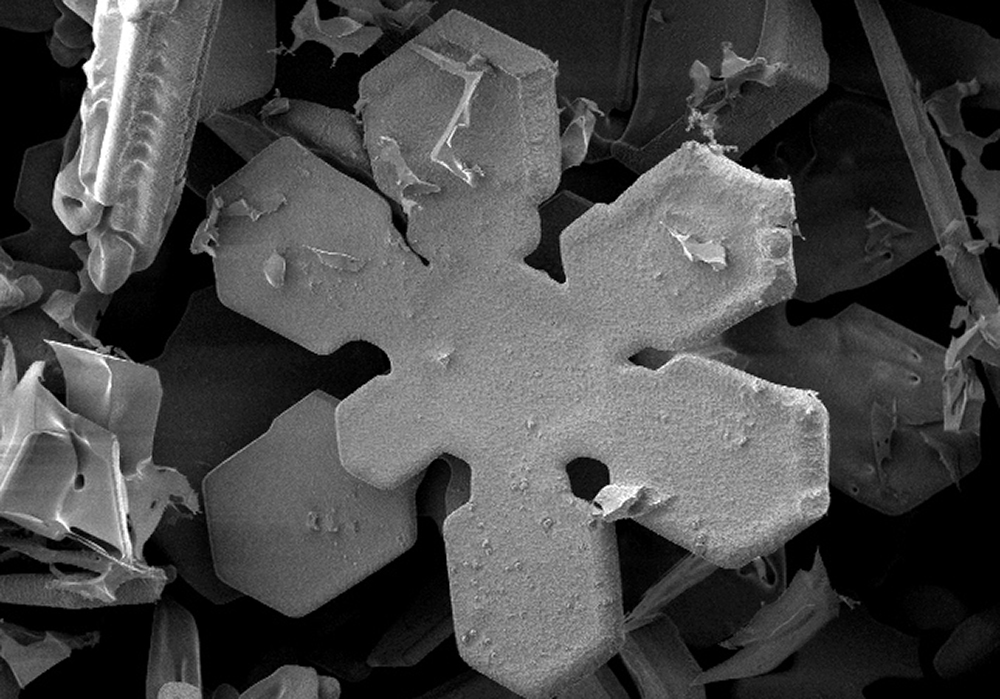
Snow
Fresh snow from Dec. 12, 2008. Hanover, NH. Columnar grain with end caps. Detail showing sublimation of flake.
 Fresh snow from Dec. 12, 2008. Hanover, NH. Flat stellar plates.
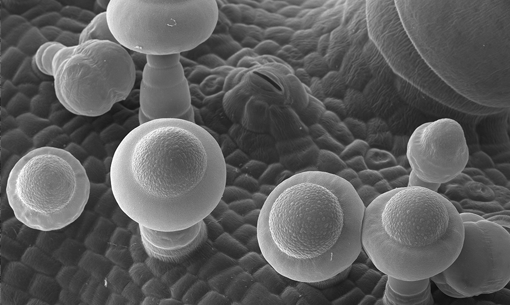
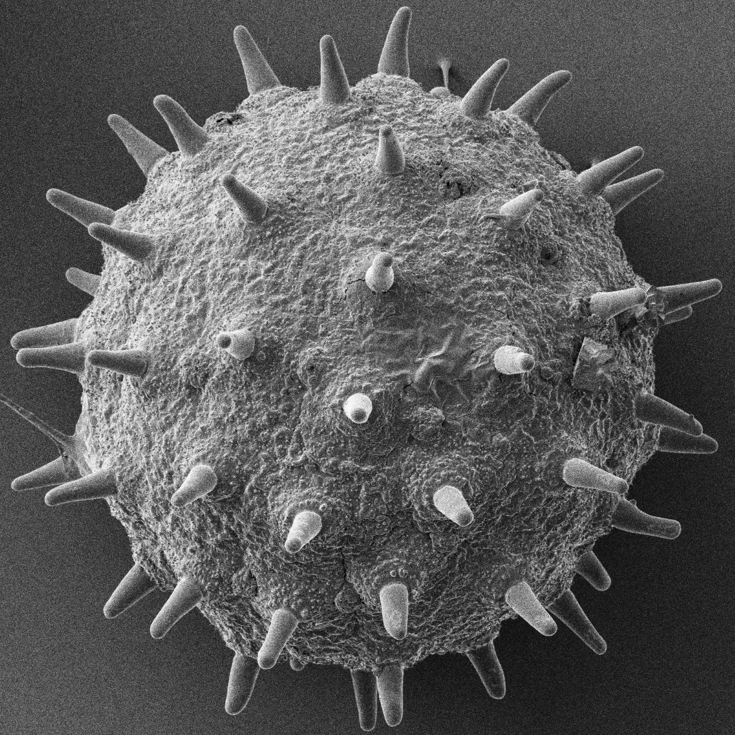
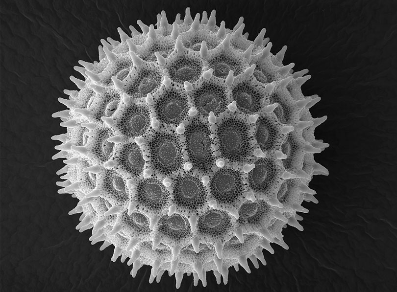
Morning Glory Flower
A grain of pollen from Morning Glory flowers.
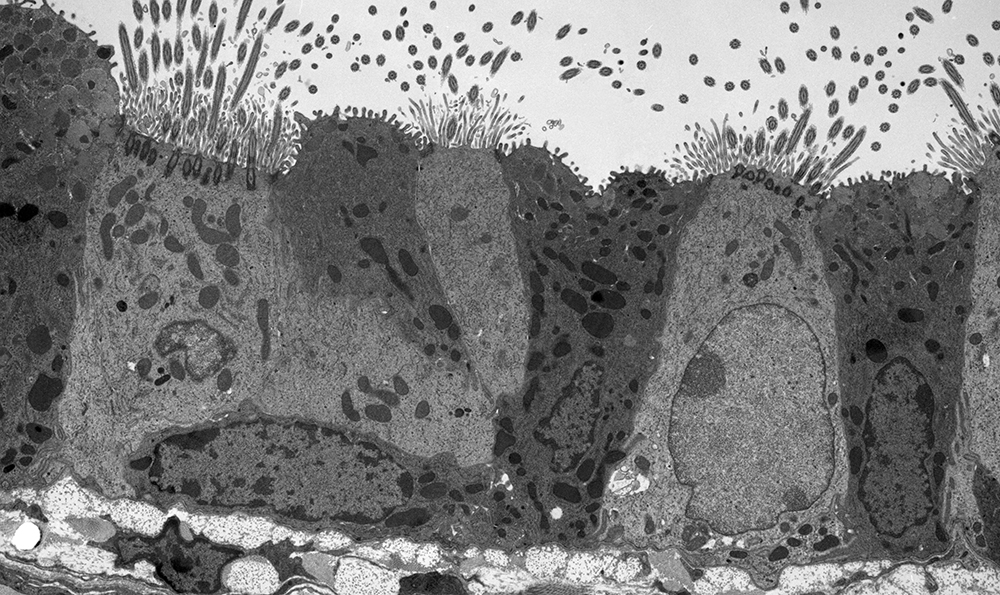
Mouse Lung
Transmission electron microscope image of a thin section cut through the bronchiolar epithelium of the lung(mouse), which consists of ciliated cells and non-ciliated cells. Image shows the ciliary microtubules in transverse and oblique section. In the cell apex are the basal bodies that are the anchoring sites for the cilia. Note the difference in size and shape between the microvilli and the cilia. At the basal end of the epithelial layer is the basement membrane. Instrument: JEOL 1010 TEM
 "Lung macrophage untreated"
 "Lung macrophage plus Asp spores"
Squash
Cucurbita maxima.